


ST1546 Anti-CBS Mouse mAb (3E1)


Produits recommandés
Aperçu
| Replacement Information |
|---|
Tableau de caractéristiques principal
| Host |
|---|
| M |
| Product Information | |
|---|---|
| Form | Liquid |
| Formulation | In PBS, pH 7.2. |
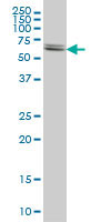
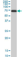
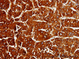
| Positive control | HeLa cells, MCF-7 cells, Human hepatocellular carcinoma tissue |
| Biological Information | |
|---|---|
| Immunogen | A recombinant polypeptide corresponding to amino acids 1-101 of human CBS, expressed as a GST fusion protein |
| Clone | 3E1 |
| Host | Mouse |
| Isotype | IgG2a |
| Physicochemical Information |
|---|
| Dimensions |
|---|
| Materials Information |
|---|
| Toxicological Information |
|---|
| Safety Information according to GHS |
|---|
| Safety Information |
|---|
| Product Usage Statements |
|---|
| Packaging Information |
|---|
| Transport Information |
|---|
| Supplemental Information |
|---|
| Specifications |
|---|
| Global Trade Item Number | |
|---|---|
| Référence | GTIN |
| ST1546 | 0 |
Documentation
Références bibliographiques
| Aperçu de la référence bibliographique |
|---|
| Hennig, B., and Diener, M., 2009. Br. J. Pharmacol. 158, 1263. Lee, M., et al. 2009. Neurobiol. Aging 30, 1523. Martin, G,R., et al. 2009. Dig. Liver Dis. 42, 103. Wallace, J,L., et al. 2009. Gastroenterology 137, 569. Xu, G,Y., et al. 2009. Mol. Pain 5, 44. Majtan, T., et al. 2008. J. Biol. Chem. 283, 34588. Wallace, J,L., et al. 2007. FASEB J. 21, 4070. Schicho, R., et al. 2006. Gastroenterology 131 1542. |