


DR1047 Sigma-AldrichAnti-UHRF1 Mouse mAb (3A11)
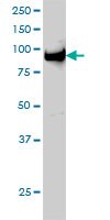

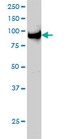
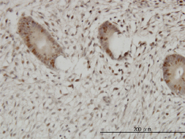
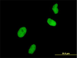
This Anti-UHRF1 Mouse mAb (3A11) is validated for use in ELISA, Immunoblotting, Immunocytochemistry, Paraffin Sections for the detection of UHRF1.
More>> This Anti-UHRF1 Mouse mAb (3A11) is validated for use in ELISA, Immunoblotting, Immunocytochemistry, Paraffin Sections for the detection of UHRF1. Less<<Anti-UHRF1 Mouse mAb (3A11) : FDS (Fiches de données de sécurité), certificats d’analyse (CoA) et de qualité (CoQ), dossiers, brochures et autres documents disponibles.
Synonymes: Anti-Ubiquitin-Like PHD/RING Finger Domain 1
Aperçu
| Replacement Information |
|---|
Tableau de caractéristiques principal
| Species Reactivity | Host | Antibody Type |
|---|---|---|
| H | M | Monoclonal Antibody |
Prix & Disponibilité
| Référence | Disponibilité | Conditionnement | Qté | Prix | Quantité | |
|---|---|---|---|---|---|---|
| DR1047-100UG |
|
100 μg |
|
— |
| References | |
|---|---|
| References | Mistry H, et al. 2008. Biochem. Biophys. Res. Commun. 375 321. |
| Product Information | |
|---|---|
| Form | liquid |
| Formulation | In PBS, pH 7.2. |
| Positive control | HeLa cells, Human endometrium tissue |
| Preservative | None |
| Quality Level | MQ100 |
| Physicochemical Information |
|---|
| Dimensions |
|---|
| Materials Information |
|---|
| Toxicological Information |
|---|
| Safety Information according to GHS |
|---|
| Safety Information |
|---|
| Product Usage Statements |
|---|
| Packaging Information |
|---|
| Transport Information |
|---|
| Supplemental Information |
|---|
| Specifications |
|---|
| Global Trade Item Number | |
|---|---|
| Référence | GTIN |
| DR1047-100UG | 04055977225969 |
Documentation
Anti-UHRF1 Mouse mAb (3A11) FDS
| Titre |
|---|
Références bibliographiques
| Aperçu de la référence bibliographique |
|---|
| Mistry H, et al. 2008. Biochem. Biophys. Res. Commun. 375 321. |