Décalcification
Decalcification Reagents: Color-Coded to Avoid Errors |
 Decalcifiers are used to prepare bone and hard tissue for microscopic analysis. The choice of reagent depends on the hardness of the material, the diagnostic purpose and the subsequent analysis. Decalcification time is based on the size and structural density of the hard tissue, as well as the composition of the decalcifying solution.
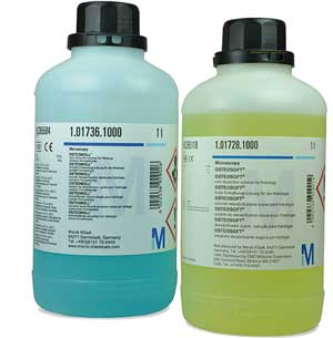
For error-free decalcification, Merck’s microscopy portfolio includes two outstanding reagents: OSTEOMOLL® and OSTEOSOFT®. These ready-to-use decalcifiers are color-coded, hence they are easily distinguishable from other laboratory reagents. Thanks to stringent quality control and excellent batch-to-batch consistency, both solutions are IVDs and CE certified. Thus, they are perfectly suited for use in diagnostic laboratories or for pharmaceutical research. Furthermore, a variety of package sizes is available to support you in managing greater demands.
Product Range
- Blue-coded OSTEOMOLL® rapid decalcifier solution for hard tissue
- Yellow-coded OSTEOSOFT® mild decalcifier solution
- Color-coded solutions prevent laboratory errors
- Reliable, reproducible results thanks to excellent quality and batch-to-batch consistency
- All products are IVDs and CE certified
- Suitable for clinical diagnostics and pharmaceutical research
- Variety of package sizes to suit demands
- Long shelf-life at room temperature
OSTEOMOLL® and OSTEOSOFT®
Hard material, like bone, teeth or cartilage, should be decalcified prior to processing. We offer two color-coded solutions for this purpose. The blue OSTEOMOLL® is used for more robust bone material and allows fast and reliable decalcification. It is an acidic formaldehyde-containing solution, which enables decalcification and fixation in one step. The yellow OSTEOSOFT® is for specimens which need gentle treatment. It preserves cell and antigen structures and allows immunohistochemical methods to be applied afterwards.


